Advanced Diagnostic Equipment

Zeiss GDx
The Zeiss GDx exam. Helping your doctor detect glaucoma—while there's still time.
What is glaucoma?
Over two million Americans have glaucoma,1 making it one of the biggest causes of legal blindness in the United States. Glaucoma can rob people of their vision even though they don't have any visual symptoms or pain. In fact, half of those with glaucoma don't even know it.2 The disease is not easily diagnosed. For example, the common "puff test," which measures eye pressure, fails to uncover glaucoma in one third of patients with the disease.3 No wonder glaucoma is called the "sneak thief of sight."
Don't let glaucoma sneak up on you.
Now there is a revolutionary new technology that can help doctors find glaucoma earlier, while there's still time: the Zeiss GDx glaucoma exam, from a trusted leader in innovative diagnostic instruments for eyecare.
What makes the Zeiss GDx exam so revolutionary?
Unlike the puff test, the Zeiss GDx exam actually lets your doctor see the pattern and thickness of the nerve fibers in the back of your eyes, then compares the results to normal values. If your nerve fibers are thinner than normal, this could indicate glaucoma long before any vision has been lost. As a result, your doctor will have more time to treat the disease.
How does the Zeiss GDx exam work?
The test is a quick and comfortable part of a complete eye exam. Plus, you don't have to have your pupils dilated. You simply look into the Zeiss GDx system while it safely scans the back of your eye. Total exam time usually takes less than a minute, and the system creates easy-to-read images that your doctor can quickly analyze.

QuantifEYE
QuantifEYE is a new technology that measures the relative health of your macula. Your Eye Care Professional can now better determine your risk for Age Related Macular Degeneration (AMD) simply by determining other risk factors and getting your MPOD measurement. Prior to this exam, you will be asked questions to assess what other risk factors you may have that can be associated with developing AMD.
The test using the QuantifEYE device is simple, quick, non-invasive, and does not require dilation prior to examination. A base-line measurement is taken, and the results are your MPOD score. If you happen to test low (low MPOD scores=increased risk for AMD) to medium, your Eye Care professional may recommend supplementation to restore depleted levels of zeaxanthin and lutein in your macula. (With MPOD testing and supplemental intervention, it is possible to increase the pigment levels in your macula, thus lowering your risk for vision loss.) Your Eye Care Professional will then be able to determine through follow up screenings if nutritional intervention is resulting in increased macular pigment.
Ask for this important test today. It's not too early to take steps to preserve your vision and restore your eyes to a healthier state.

Digital Retinal Imaging
Our ability to view your internal eye health is now dramatically improved with retinal imaging, providing assistance with the early detection of eye diseases (pre-cancerous and cancerous lesions, diabetic retinopathy, AIDS related retinopathy, optic nerve disease, macular degeneration, retinal detachments, etc.)
Many serious eye diseases are painless, symptom-free, and slowly progressive. These conditions can often continue undetected for many years, potentially resulting in significant vision loss or even blindness. Early diagnosis is essential.
Additionally, systemic diseases such as high blood pressure and diabetes can be detected during a retinal exam. Digital Retinal Imaging creates a permanent record and baseline for your medical file, which gives your doctor comparisons for tracking and diagnosing potential eye disease. The doctor will review your images with you today.

Computerized Corneal Topography
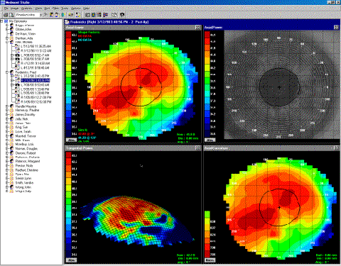
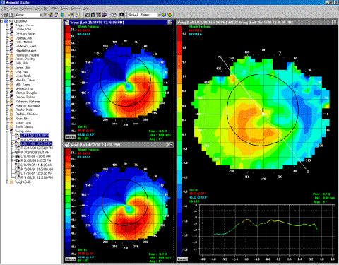
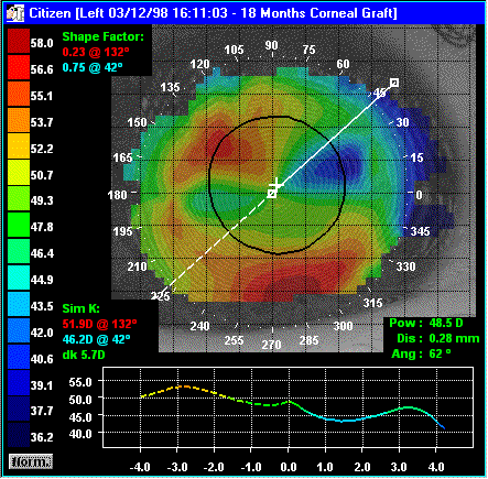
Of all the technology currently available, corneal topography provides the most detailed information about the curvature of the cornea. Using a very sophisticated computer and software, thousands of measurements are taken and analyzed in just seconds.
The computer generates a color map from the data. This information is useful to evaluate and correct astigmatism, monitor corneal disease, and detect irregularities in the corneal shape. The map is interpreted much like other topography maps. The cool shades of blue and green represent flatter areas of the cornea, while the warmer shades of orange and red and represent steeper areas.
This corneal map allows the physician to formulate a "3-D" perspective of the cornea's shape. Measuring astigmatism is important for planning refractive surgery, fitting contact lenses, and calculating intraocular lens power.


Zeiss OCT
Vonnahme Eye Care can now provide an even more thorough examination and diagnosis of your retina than ever before with the Zeiss Stratus OCT. This cutting-edge diagnostic system allows our doctors to get a complete cross-sectional view of your retina using an optical measurement known as low-coherence interferometry.
The technology is much like ultrasound technology, except that it uses light rather than sound.
The Zeiss Stratus OCT scans an infrared light across the retina, which generates a cross-sectional image of the tissue in microscopic detail. It generates a measurement of tissue and distance of up to 1/100 of a millimeter and over 500,000 data points.
This allows us to view your retina as if it were under a microscope, enhancing our ability to diagnose and manage a wide-range of retinal disorders, including diabetic retinopathy, macular degeneration, ocular histoplasmosis, and macular holes.

